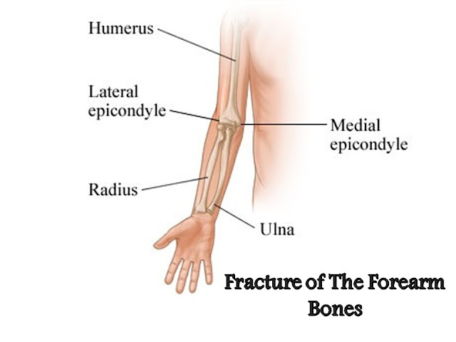
Fracture of the forearm bones produces considerable problems. The greenstick fracture in children can be reduced easily whereas fracture in adults may at times require open reduction and the open reduction or surgery is done with the help of orthopedic instruments by the orthopedic surgeons. Rotational and angulatory deformities are likely to impair the activity of the limb even after the union of the fracture.
Certain important facts must be understood properly in order to deal with lesions of the forearm bones.
Anatomical Factors: The ulna is supposed to be the continuation of the humerus whereas the radius is the continuation of the hand. The ulna mainly participates in the stability of the elbow-joint. Fracture of one bone is therefore associated with dislocation of the unstable joint formed by the other. These unstable joints are formed at the upper part by the head of the radius with the humerus, while at the lower end by the distal end of the ulna with the carpal bones.
Muscular Attachments: The three main muscles of supination and pronation, e.g., pronator teres, pronator quadratus and supinator muscles tend to displace the fractured segments in the case of fracture of the radius. The amount of displacement depends upon the site of the lesion.
(a) Fracture of the upper third of radius: In lesion at the upper third of radius, the fracture line passes above the insertion of pronator teres. The proximal radial segment remains supinated by the action of the supinator muscle while the distal segment remains pronated by the action of the pronator teres and pronator quadratus muscles. The stable reduction can only be achieved by immobilizing the forearm in the supinated position.
(b) Fracture of the mid-third of radius: In cases of fracture below the insertion of the pronator teres, the stability is obtained with the forearm being immobilized midway between prone and supine positions.
(c) Fracture of the distal third of radius: The distal segment remains pronated by the unopposed action of the pronator quadratus. The proximal segment maintains the mid-prone position by the combined action of the pronator teres and supinator muscles. The forearm should be immobilized in the prone position which enables the proximal segment to remain aligned with the distal one.
Alignment of fracture: Presence of rotational or angular deformity interferes with the important pronation and supination movements. Failure to reduce the fracture by closed technique will require operative correction. The functional ability of the forearm bone must be recognized, mere union of the fracture is not enough. One should contemplate producing a normal functioning upper limb after the union has been restored.
Anatomical Landmarks:
I. By palpation: The proximal part of the forearm bones are covered by more muscles than the distal end and is therefore difficult to palpate properly. The posterior border of the ulna is subcutaneous and can be felt easily. The manipulator takes the advantage of these landmarks during the process of reduction. By simple palpation, one can feel the aligned position before plaster is applied.
II. Radiology: The bicipital-tuberosity of the radius is an important landmark. The amount of rotation the proximal part of radius undergoes after fracture can be assessed by comparing the bicipital tuberosity of the normal side with the aid of x-ray.


